Breakthrough

On a hot afternoon in late summer 2010, a man in his 30s drove an all-terrain vehicle on an unpaved path. He was doing nearly 40 miles per hour on rough terrain. And though he was strong—a construction worker by trade—his ATV hit a bump for which he wasn’t prepared.
He drove off the road. The airborne ATV flipped end-over-end and the man flew into the brush. He wore no helmet and his head hit the jagged earth hard. It took his riding companions more than a minute to reach their unconscious friend and nearly 30 minutes to get to a phone to call an ambulance.
Medics transported him via life flight helicopter to UPMC Presbyterian in Oakland, where Dr. David Okonkwo, a 37-year-old brain trauma specialist, worked to keep him alive. After observation and a few preliminary tests, the doctor knew his patient would survive the first 24 hours, but he didn’t know much else. That was not unusual.
Between 2002 and 2006, the Center for Disease Control and Prevention estimated that there were 52,000 deaths, 275,000 hospitalizations and more than 1.3 million emergency room visits in the U.S. related to traumatic brain injury. In more than 90 percent of the cases, doctors used Magnetic Resonance Imaging (MRI), Computed Tomography (CT) and other scanning technologies and found very little. Through magnetic and radio frequency fields, MRIs produce black and white images of the human interior that are much clearer than CT scans or traditional X-rays. However, with brain scans, MRIs are limited. They help doctors locate the injury but are imprecise in determining its extent.
The young man in the ATV accident declined to be interviewed for this article, and because of confidentiality rules, some details of his treatment have been concealed, including his real name. We’ll call him Joe. Six days into his ordeal, Joe underwent his first MRI. It showed that he had a bruised brain. In much of the body, a bruise causes discomfort but eventually heals. A brain bruise, however, means part of the brain has died, leading to anything from temporary slurred speech to a persistent vegetative state or death. Dr. Okonkwo didn’t know what that bruise would mean for Joe’s future. For two weeks, Joe remained comatose, and his family worried that he might never awaken. “In many ways,” Dr. Okonkwo said, “we were flying blind.”
Dr. Okonkwo, however, knew where to turn. Three months earlier, he sat in on a short presentation by Walter Schneider, a University of Pittsburgh psychology professor and senior scientist at Pitt’s Learning Research and Development Center. Schneider and his colleagues at Pitt and UPMC are developing a brain-scanning technology that may allow doctors to determine not only where brain injuries are, but also how the injuries will influence a patient’s ability to speak, walk and think.
For decades, Schneider had mapped regions of the brain using MRIs. He had published results of various experiments in scholarly journals and books on topics such as human vision and “the architecture of language processing.” But his latest project was not merely a regional map; it was a plan to create an atlas of the human brain, complete with 250,000 fiber pathways detailing the brain’s structure and its means for communication with the body.
What Schneider promised was not only a deeper understanding of the brain but also, in essence, a new and superior form of MRI—a scan that would help a neurosurgeon understand how a brain bruise might affect neurological connections and affect a patient’s life.
“It took me about 60 seconds to know that this was something I needed to be a part of,” Dr. Okonkwo said. “I knew this was a paradigm shift that could change everything.”
At 6 feet, 2 inches tall, the lanky 61-year old Schneider sports a full head of short, gray hair. In a slightly faded button-down shirt, khakis and lace-up leather sandals, he is a portrait of learned relaxation. His sixth-floor Oakland office is crammed with books, papers and a computer with two monitors that display colorful images—occasionally of the inside of his own brain. The psychology professor dabbles in education, cognition and radiology, among other things. When pressed, he’ll tell you he’s an investigator.
For more than 30 years, he’s been probing the way we think and how brain activity influences cognition, movement and speech. For years, he’s wanted to create technology that would map the connections of the human brain for clinical use and basic research. So about three years ago, he and his small staff started looking into technology called Diffusion Tensor Imaging (DTI), a technology developed in the early 1990s that had gained traction at the National Institutes of Health.
“DTI had the right intention,” Schneider said, smiling.
What DTI offered that an MRI could not was the ability to see which direction water was diffusing within the human body. And if a brain scan can determine where and how water is traveling within the body, it can help doctors determine why and for what purpose. “DTIs gave the doctor the ability to say, ‘This water is going up; this water is going forward; this water is going laterally.’ ”
But DTI had nowhere near the acceptable accuracy for an effective brain scan, Schneider said.
Doctors had to connect the dots from water source to water destination. It was no easy task, involving long hours and questionable results. In order to get the full picture without connecting the dots, they needed much higher resolution and accuracy than DTI offered in the early 1990s. By 2009, the technology had developed. So in March that year, Schneider his staff organized a worldwide contest.
Dubbed the Pittsburgh Brain Competition, the project convened the world’s foremost neurological experts and offered a $10,000 prize for improving DTI resolution. The winning teams—one from St. Louis and one from the Netherlands—produced their results. And from there, as Schneider said, “we in Pittsburgh were able to actually replicate those two leading techniques and then go beyond what they did.”
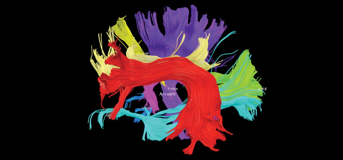
The result is High Definition Fiber Tracking, which makes the brain’s connections visible. Instead of a flat, black and white MRI image, the new technology shows a three-dimensional image of the brain filled with what looks like thin, multi-colored strands of spaghetti. These strands, however, are more like fiber optic cables than food. And they can be mapped to show doctors which brain connections are functional and which aren’t.
Schneider believes the technology can help revitalize surgical planning and operating room guidance. “If you know where the cables are going, how they connect and what function they serve, you’re less likely to cut something that’s going to cause irreparable damage.” He also believes High Density Fiber Tracking will change the way doctors treat genetic brain disorders such as autism, as well as traumatic brain injuries such as the one Joe experienced when he wrecked his ATV.
“It really does present tremendous opportunities,” Schneider said. With Dr. Okonkwo’s help, Schneider and his team typically look into two clinical cases per week—one in neurosurgery, one in traumatic brain injury. Cases are chosen on the basis of their complexity and whether UPMC neurosurgeons are having trouble with diagnosis or surgery.
Joe had been in a coma for two weeks before he regained consciousness. After the initial joy, though, he realized he couldn’t move the left side of his body. Another MRI was inconclusive. So his family and doctors discussed next steps. Schneider was not involved in the discussion but added, “If that was the brain of a loved one, I’d sure want them to use that technology.”
Joe began rehabilitation within a month of his injury—a minimum of three hours per day of physical, speech and occupational therapy. It began with the basics: using a fork, feeding himself safely, learning to walk and talk again. After a month, progress was slow, and neither Joe nor his family knew if this was the result of his not working hard enough or other factors. Uncertainty is common with brain injury. Friends and family often wonder whether the patient is malingering, even whether the injury is real. It adds another element to the frustration of recovery.
Joe and his family ultimately agreed, and Dr. Okonkwo secured funds and permission, to run a High Density Fiber Tracking scan on Joe’s brain.
It was not a fairy tale ending. The scan showed that about 54 percent of the connections in Joe’s brain influencing movement of his left leg were severed and likely dead. At best, in other words, he would have to make do with about half the strength of his left leg prior to his injury. The scan showed that about 97 percent of the connections influencing movement of his left arm were severed and likely dead. At best, his arm would operate at about 3 percent strength; in other words, it would be largely immobile for the rest of his life.
“Is he happy with his situation? No,” Dr. Okonkwo said. “It’s wonderful that he’s recovered as much as he has. But, when you’re well, you just assume you’re going to be well every day of your life. We take that stuff for granted. So there’s still frustration associated with the things he can’t do and the functional limitations he has. But at least now he some idea about what he can expect.
“Now, rather than spending most of his days being angry about not knowing, he understands what he has to work with and how he can deal with it. The hardest part really is the unknown. And anything we can do to minimize that is a victory for everyone.”


